
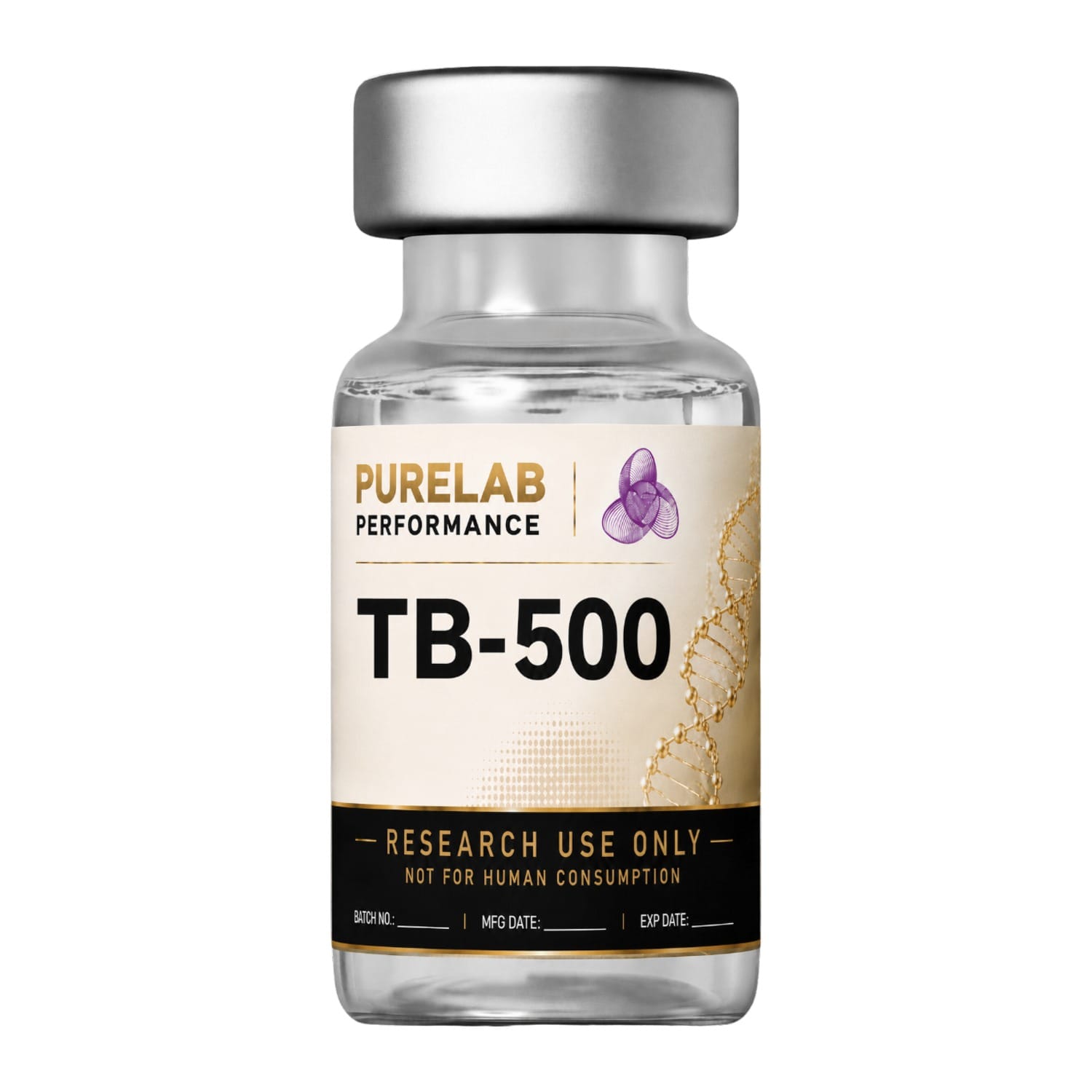
What TB-500 actually is
This is the first thing most vendors get wrong, so we will start here.
TB-500 is not thymosin β4. Thymosin β4 (Tβ4) is a 43-amino-acid protein found throughout mammalian tissue. TB-500 is a synthetic seven-amino-acid fragment of it — residues 17 to 23, the actin-binding domain, with an acetyl group added to the N-terminus. Analytical chemists who characterised commercial TB-500 preparations by high-resolution mass spectrometry identified the active ingredient as Ac-LKKTETQ[1].
The distinction matters for two reasons. First, most of the animal work people cite when discussing “TB-500” was performed with full-length Tβ4 — a different molecule, with a different mass and a different pharmacokinetic profile, and forty-three residues instead of seven. Second, the safety literature discussed below is a literature about Tβ4, and whether it transfers to a seven-residue fragment is an open question that nobody has answered.
Structure and identity
Every batch we supply is assayed by an independent laboratory using HPLC-UV-MS. The mass spectrum is compared against an authentic reference standard to confirm molecular identity; the peptide’s peak area is measured against the total chromatogram area to establish chromatographic purity; and total peptide mass in the vial is measured against the stated label claim.
Measured purity for the current lot is Certificate pending. We publish the certificate in full when the analysis returns, and it can be verified with the issuing laboratory independently of us. We do not print a purity number before we have one.
Where the evidence stands
This is the section most vendors skip. On this compound it is by some distance the most important one on the page.
Actin sequestration, cell migration, ILK/AKT/β-catenin signalling — largely with full-length Tβ4Extensive
Rodent tumour models; equine pharmacokinetic and detection work with the actual heptapeptideModerate
Tβ4 overexpression associated with invasion, EMT and metastasis — independent groups, gain- and loss-of-functionConsistent
Not approved for human therapeutic use by FDA or any other regulatorNone
The oncology safety signal in vitro rodent
This section is the reason this page reads the way it does. The finding is not hypothetical, and it is not one lab’s outlier: it is a coherent body of work from independent groups on three continents, using both gain-of-function and loss-of-function designs, and it points consistently in one direction.
Overexpression increases invasiveness and MMP-7
In a 2004 study in Oncogene, SW480 human colon carcinoma cells were stably transfected to overexpress Tβ4. The Tβ4-overexpressing cells showed a marked increase in invasiveness, accompanied by increased levels and enzymatic activity of matrix metalloproteinase-7 (MMP-7) — an enzyme that degrades basement membrane and is central to how tumour cells breach tissue boundaries. Levels of Fas fell significantly, and the cells became less susceptible to Fas-ligand-mediated apoptosis. In matched patient samples, Tβ4 mRNA, β-catenin, c-Myc and MMP-7 were all higher in metastatic liver lesions than in the primary colorectal tumours they came from[2].
An earlier report from the same group had already shown that forcing Tβ4 expression in SW480 cells increased growth rate, colony formation in soft agar and motility, while downregulating E-cadherin at intercellular junctions[3].
Overexpression triggers epithelial-mesenchymal transition
A 2006 paper in Oncogene reported that SW480 cells overexpressing Tβ4 adopted a scattering, fibroblastic morphology with reduced intercellular contacts, lost E-cadherin, and accumulated cytosolic β-catenin — the two most prominent markers of epithelial-mesenchymal transition (EMT), the program by which epithelial tumour cells acquire the capacity to invade and metastasise. The mechanism ran through upregulation of integrin-linked kinase (ILK)[4]. A 2014 study in Biochemical and Biophysical Research Communications independently confirmed the ILK/AKT/β-catenin axis, and reported that Tβ4 was significantly overexpressed in colorectal cancer tissue relative to adjacent normal tissue, with high levels correlating with disease stage[5].
Histologically the picture is consistent. Across 76 colorectal adenocarcinomas, Tβ4 immunoreactivity increased progressively from the superficial toward the deepest tumour regions, and the strongest expression was found in invading tumour cells at the invasion front, matched by a progressive loss of E-cadherin in those same cells[6].
Higher expression, worse outcomes
In tissue from 143 colorectal cancer patients, high Tβ4 expression was significantly correlated with lymphovascular invasion, invasion depth, regional lymph node metastasis, distant metastasis and TNM stage. On multivariate analysis, patients with high Tβ4 expression showed poorer recurrence-free survival (p = .001) and poorer overall survival (p = .005)[7].
Knocking it down reduces metastasis
The converse experiment has also been done, and it is the most direct of the three. In a 2015 study in Oncotarget, Tβ4 expression in B16F10 murine melanoma cells was disrupted using TALEN gene editing. In a murine model, inhibition of Tβ4 expression significantly decreased lung metastasis of B16F10 cells[8]. A separate study using lentiviral shRNA against Tβ4 found that tumour cell migration decreased when Tβ4 was silenced, and increased again when Tβ4 protein was added back[9].
These are studies of endogenous Tβ4 expression inside tumour cells — not studies of exogenously administered TB-500 in a healthy organism. That is a real difference and we are not going to overstate it. But the mechanism implicated — actin remodelling driving cell migration, ILK/AKT/β-catenin signalling, E-cadherin loss, MMP-7 induction — is precisely the mechanism by which this peptide family is claimed to promote repair. The property that makes a cell migrate into a wound is the property that makes a cell migrate out of a tumour. No study has established that this concern is misplaced, because no study has gone looking.
Mechanism at the cell in vitro
Tβ4 is the major actin-sequestering protein in mammalian cells: it binds monomeric G-actin and modulates the assembly and disassembly of F-actin microfilaments[3]. The LKKTETQ heptapeptide that constitutes TB-500 is the actin-binding domain within that protein, which is the stated rationale for using the fragment rather than the whole molecule[1].
Downstream, Tβ4 has been reported to regulate Rap1- and Rac1-GTPase activity under hypoxia/reoxygenation conditions[8], to act together with HIF-1α in driving tumour cell migration[9], and to negatively regulate GSK-3α, β-catenin and E-cadherin via ERK phosphorylation[10]. Every one of these is a cell-culture finding. None of them is a finding about a living human.
Repair and regeneration models preclinical
The commercial claims made for TB-500 — that it promotes endothelial cell differentiation, angiogenesis in dermal tissue, keratinocyte migration and collagen deposition, and decreases inflammation — were catalogued by a racing laboratory in the course of developing a detection assay, which described them explicitly as claims made for the veterinary preparation rather than as established findings[11]. We reproduce that framing because it is the accurate one.
We are not aware of a randomised, controlled study of the Ac-LKKTETQ heptapeptide in any repair model, in any species, that meets a modern standard of evidence. If such a study exists, we would genuinely like to see it, and we will update this page.
The human data — all of it human
There is none.
There are no published clinical trials of TB-500 (Ac-LKKTETQ) in humans — not a randomised trial, not a pilot study, not a case series. The only human-relevant analytical work is a doping-control method development paper, which synthesised the peptide by solid-phase synthesis and established an approach for detecting it in human plasma and urine[1]. That is a paper about detecting the compound in people who have used it. It is not a paper about whether it does anything, or whether it is safe.
Long-term safety in humans is entirely uncharacterised. Read alongside the oncology literature above, that gap is not a neutral fact.
Limitations of the literature
The molecule in the papers is not the molecule in the vial. The regeneration literature is overwhelmingly about a 43-residue protein. The product is a 7-residue fragment. These are routinely conflated by people selling the fragment, and the conflation consistently flatters the fragment.
The safety literature is the stronger literature. This is unusual, and worth saying plainly. The oncology findings above come from independent groups in Taiwan, Korea, Italy and elsewhere, converge on a consistent mechanism, and include both gain-of-function and loss-of-function experiments. The efficacy claims, by contrast, rest largely on preclinical work with a different molecule.
Nobody has run the experiment you would want run. No published study has administered TB-500 to a healthy animal over an extended period and examined tumour incidence. Until someone does, the honest position is that the question is open.
Regulatory and anti-doping status
TB-500 is prohibited by the World Anti-Doping Agency by name, at all times, in and out of competition. It appears on the Prohibited List under section S2.3, Growth Factors and Growth Factor Modulators, in these words: “Thymosin-β4 and its derivatives e.g. TB-500”[12]. Substances in class S2 are non-Specified Substances, which carries the more severe sanctioning framework under the World Anti-Doping Code.
It is also detectable. A validated LC-MS method developed at a racing laboratory confirmed N-acetylated LKKTETQ down to 0.01 ng/mL in equine urine and 0.02 ng/mL in equine plasma, and identified the parent peptide together with its metabolites in post-administration samples from horses given a single 10 mg dose[11]. A parallel method for human plasma and urine was published the same year[1]. Anyone assuming this compound is invisible to testing is working from information that was already out of date in 2012.
TB-500 has not been approved for human therapeutic use by the FDA or by any other governmental regulatory health authority.
What we don’t know
- Whether the oncology signal associated with full-length Tβ4 transfers to the Ac-LKKTETQ fragment. Nobody has tested this.
- Whether administered TB-500 affects tumour incidence or progression in any species. Nobody has tested this either.
- Whether the regeneration effects attributed to Tβ4 occur with the heptapeptide at all.
- Long-term safety in humans — entirely uncharacterised.
- Per-kilogram dosing in animal models does not scale to humans, and nothing on this page should be read as implying that it does.
We supply this compound for laboratory research.
TB-500 supplied by PureLab Performance is furnished strictly for in-vitro laboratory research. It is not a medicine or a drug and has not been approved by the FDA to prevent, treat, or cure any medical condition, ailment, or disease. Bodily introduction of any kind into humans or animals is strictly forbidden by law. Per-kilogram dosing in animal models does not scale to humans. Purchasers must be 18 or older and qualified to handle research chemicals.
References
Retrieved from PubMed. DOIs link to the original publications.
- Esposito S, Deventer K, Goeman J, Van der Eycken J, Van Eenoo P. “Synthesis and characterization of the N-terminal acetylated 17-23 fragment of thymosin beta 4 identified in TB-500, a product suspected to possess doping potential.” Drug Test Anal. 2012;4(9):733–38. DOI
- Wang WS, Chen PM, Hsiao HL, Wang HS, Liang WY, Su Y. “Overexpression of the thymosin beta-4 gene is associated with increased invasion of SW480 colon carcinoma cells and the distant metastasis of human colorectal carcinoma.” Oncogene. 2004;23(39):6666–71. DOI
- Wang WS, Chen PM, Hsiao HL, Ju SY, Su Y. “Overexpression of the thymosin beta-4 gene is associated with malignant progression of SW480 colon cancer cells.” Oncogene. 2003;22(21):3297–306. DOI
- Huang HC, Hu CH, Tang MC, Wang WS, Chen PM, Su Y. “Thymosin beta4 triggers an epithelial-mesenchymal transition in colorectal carcinoma by upregulating integrin-linked kinase.” Oncogene. 2007;26(19):2781–90. DOI
- Piao Z, Hong CS, Jung MR, Choi C, Park YK. “Thymosin β4 induces invasion and migration of human colorectal cancer cells through the ILK/AKT/β-catenin signaling pathway.” Biochem Biophys Res Commun. 2014;452(3):858–64. DOI
- Nemolato S, Restivo A, Cabras T, et al. “Thymosin β 4 in colorectal cancer is localized predominantly at the invasion front in tumor cells undergoing epithelial mesenchymal transition.” Cancer Biol Ther. 2012;13(4):191–97. DOI
- Lee SY, Park MJ, Lee HK, Son HJ, Kim CN, Kim JH, Kang DW. “Increased Expression of Thymosin β4 Is Independently Correlated with Hypoxia Inducible Factor-1α (HIF-1α) and Worse Clinical Outcome in Human Colorectal Cancer.” J Pathol Transl Med. 2017;51(1):9–16. DOI
- Lee JW, Ryu YK, Ji YH, Kang JH, Moon EY. “Hypoxia/reoxygenation-experienced cancer cell migration and metastasis are regulated by Rap1- and Rac1-GTPase activation via the expression of thymosin beta-4.” Oncotarget. 2015;6(12):9820–33. DOI
- Ryu YK, Im YS, Moon EY. “Cooperation of actin-sequestering protein, thymosin β-4 and hypoxia inducible factor-1α in tumor cell migration.” Oncol Rep. 2010;24(5):1389–94. DOI
- Ryu YK, Lee YS, Lee GH, Song KS, Kim YS, Moon EY. “Regulation of glycogen synthase kinase-3 by thymosin beta-4 is associated with gastric cancer cell migration.” Int J Cancer. 2012;131(9):2067–77. DOI
- Ho ENM, Kwok WH, Lau MY, Wong ASY, Wan TSM, Lam KKH, Schiff PJ, Stewart BD. “Doping control analysis of TB-500, a synthetic version of an active region of thymosin β₄, in equine urine and plasma by liquid chromatography-mass spectrometry.” J Chromatogr A. 2012;1265:57–69. DOI
- World Anti-Doping Agency. Prohibited List — S2.3, Growth Factors and Growth Factor Modulators: “Thymosin-β4 and its derivatives e.g. TB-500.” wada-ama.org/en/prohibited-list
Reviews
There are no reviews yet.